Stress cardiomyopathy - Takotsubo
| Case description: Mimicking myocardial infarction by a stress cardiomyopathy
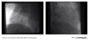
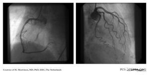
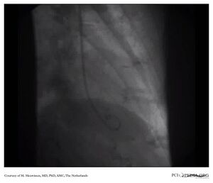
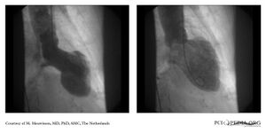
An 81-year-old woman with no cardiac history presented with acute chestpain which radiated to the left arm. The blood pressure was 140/80 mmHg and a heart rate of110/min. Physical examination revealed no abnormalities. The electrocardiogram was compatible with acute anterior myocardial infarction. (A) Immediate coronary angiography showed normal coronary arteries (B and C). A left ventricular (LV) angiogram revealed a Tako-tsubo-like cardiomyopathy, recognized by a hypercontractile base and a bulging out of the LV-apex at systole (D), which normalizes at diastole (E). This typical LV-angiogram resembles a local octopus trap in Japan , where this cardiomyopathy was first described. Although clinical presentation can be quite severe, prognosis is usually good with complete LV recovery. It occurs commonly in post-menopausal woman, usually provoked after extreme emotional stress. In a second interview, the patient told she had a severe emotional experience at a parking lot that morning, after which she developed chestpain. | |
| Courtesy of: Courtesy of M. Meuwissen, MD, PhD, AMC, The Netherlands | |
| <flash>file=MM0097.swf | <flash>file=MM0098.swf |
| Takotsubo_LCA | Takotsubo_LVangio |
| <flash>file=MM0099.swf | 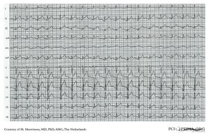
|
| Takotsubo_RCA | Electrocardiogram (A) |
|
|
| Electrocardiogram (B) and (C) | Coronary Arteries (B) and (C) |
|
|
| Coronary Arteries (D) | Left Ventricular Angiogram (D) and (E) |